Diagnostic Services
We offer the following Diagnostic Services:
CT scanning
Radiology
Nuclear medicine
MRI
EEG
Respiratory therapy
Pulmonary function testing
Clinical laboratory services
GI laboratory services
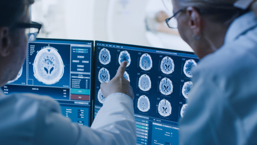
Computed Tomography (CT) Scan
What is a CT Scan?Doctor looking at ct scan results A CT scan - also called computerized tomography or just CT - is an X-ray technique that produces images of your body that visualize internal structures in cross-section rather than the overlapping images typically produced by conventional X-ray exams. Conventional X-ray exams use a stationary X-ray machine to focus beams of radiation on a particular area of your body to produce two-dimensional images on film or digital detector, much like a photograph. However, CT scanners use an X-ray unit that rotates around your body and a powerful computer. The result is a set of cross-sectional images, like slices, of the inside of your body.
CT scan of the body is performed to:
Detect and monitor diseases such as cancer or heart disease
Detect internal injuries and internal bleeding
Diagnose muscle and bone disorders, such as bone tumors and fractures
Pinpoint the location of a tumor, infection or blood clot
Diagnostic X-Ray
An X-ray is a quick, painless test that produces images of the structures inside your body - particularly your bones. X-ray technology is used to examine many parts of the body.BONES x-rays:
Arthritis
Bone cancer
Fractures and infections
Osteoporosis
CHEST:
Blocked blood vessels
Cancer
Enlarged heart
Lung infections or conditions
ABDOMEN:
Digestive tract problems
Modified barium swallows
Other fluoroscopy procedures
Swallowing Evaluation - used to examine the upper GI (gastrointestinal) tract which includes the esophagus and part of the stomach
Swallowed items
Magnetic Resonance Imaging (MRI)
What is MRI of the Body?Magnetic Resonance Imaging (MRI) is a noninvasive medical test that helps physicians diagnose and treat medical conditions.
MRI uses a powerful magnetic field, radio frequency pulses and a computer to produce detailed pictures of organs, soft tissues, bone and virtually all other internal body structures. The images can then be examined on a computer by a radiologist. The images can be stored and copied on a CD. The MRI does not use ionizing radiation (x-rays).
Detailed MR images allow physicians to better evaluate various parts of the body and certain diseases that may not be assessed adequately with other imaging methods such as x-ray, ultrasound or computed tomography.
MRI of the body is performed to evaluate:
Blood Vessels
Breasts
Organs of the chest and abdomen - including the heart, liver, biliary tract, kidney, spleen, and pancreas and adrenal glands
Pelvic organs including the reproductive organs in the male (prostate and testicles) and the female (uterus, cervix and ovaries)
Physicians use the MRI examination to help diagnose or monitor treatment for conditions such as:
Blockages, or enlargements of blood vessels, including the aorta, renal arteries, and arteries in the legs
Breast cancer and implants
Causes of pelvic pain in women, such as fibroids, endometriosis and adenomyosis
Certain types of heart problems
Cysts and solid tumors in the kidneys and other parts of the urinary tract
Diseases of the liver, such as cirrhosis and that of other abdominal organs, including the dile ducts, gallbladder, and pancreatic ducts
Suspected uterine congential abnormalities/anomalies in women undergoing evaluation for infertility
Tumors and other abnormalities of the reproductive organs (e.g. uterus, ovaries, testicles, prostate)
Tumors of the chest, abdomen, or pelvis
For useful advice about X-rays, MRIs and other medical imaging tests, click here!